- Home/
- SSC & Railways/
- SSC CGL/
- Article
Human Circulatory System – Organs, Diagrams, Functions & Notes
By BYJU'S Exam Prep
Updated on: September 25th, 2023

The Human Circulatory System is a vital part of the human body. It is a complex network of organs and vessels responsible for the transportation of blood, oxygen, and nutrients throughout the body. It includes the heart, blood vessels (arteries, veins, and capillaries), and blood. The circulatory system plays a crucial role in maintaining overall health by regulating body temperature, and protecting us against infections and diseases.
The circulatory system is divided into two types: open circulatory systems and closed circulatory systems. Humans have a closed circulatory system in which blood flows through a closed network of blood vessels, while in an open circulatory system, blood flows through open spaces. Let’s learn more about Human Circulatory System and its components in detail.
Table of content
-
1.
Human Circulatory System

-
2.
Human Circulatory System Notes PDF
-
3.
Human Circulatory System Diagram
-
4.
Organs of Human Circulatory System
-
5.
Features of Human Circulatory System
-
6.
Functions of Human Circulatory System
-
7.
Human Circulatory System: Working
-
8.
Human Circulatory System: Important Points
Human Circulatory System
Human Circulatory System is the system of organs including heart, blood vessels and blood which is circulated throughout the human body.
The human body is a complex system that relies on a number of processes to function effectively. To ensure that these vital operations run well, the various regions of the body must be provided with the essential components. William Harvey was the one who discovered the circulatory system.
Important |
|
SSC CHSL 2023 Tier 1 Exam Analysis – Feedback Form Correct Questions सटीक Analysis के साथ, अपना Exam Experience हमारे साथ share करें Fill out the Form Now>> https://forms.gle/VMABszUvdJgnBgnn7 |

Human Circulatory System Notes PDF
Having human circulatory system notes in a PDF format is important candidates planning to appear for the upcoming SSC exams or other competitive exams. The circulatory system is a complex system, and having a well-organized and easy-to-understand set of notes can help aspirants learn the concepts effectively.
> Human Circulatory System Notes PDF
Human Circulatory System Notes PDF also makes it easy to access the notes on any device and take them on the go. It can also be helpful for revision and exam preparation, as students can quickly search for specific topics and review the material at their own pace. Interested candidates can download the Human Circulatory System notes PDF from the direct link mentioned here.
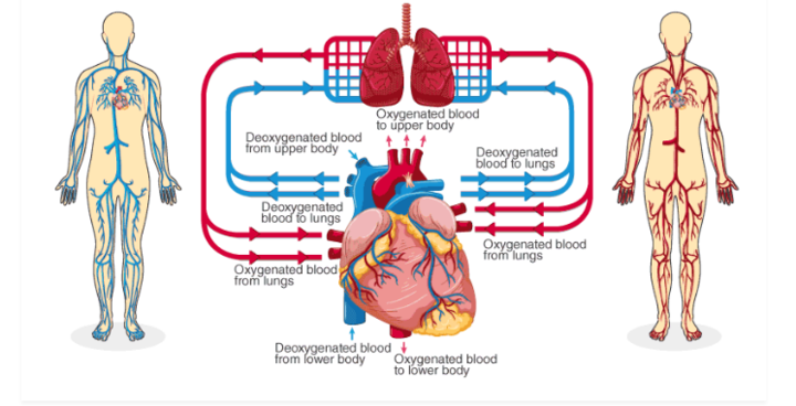
Human Circulatory System Diagram
The human circulatory system transports necessary nutrients and minerals throughout the body as well as metabolic waste products out from the body. A well-labeled circulatory system diagram is shown below.
Organs of Human Circulatory System
There are 4 main organs of the Human Circulatory System that carry out all the functions. These vital organs are as follows:
- Lymphatic System
- Blood Vessels
- Blood
- Heart
Heart
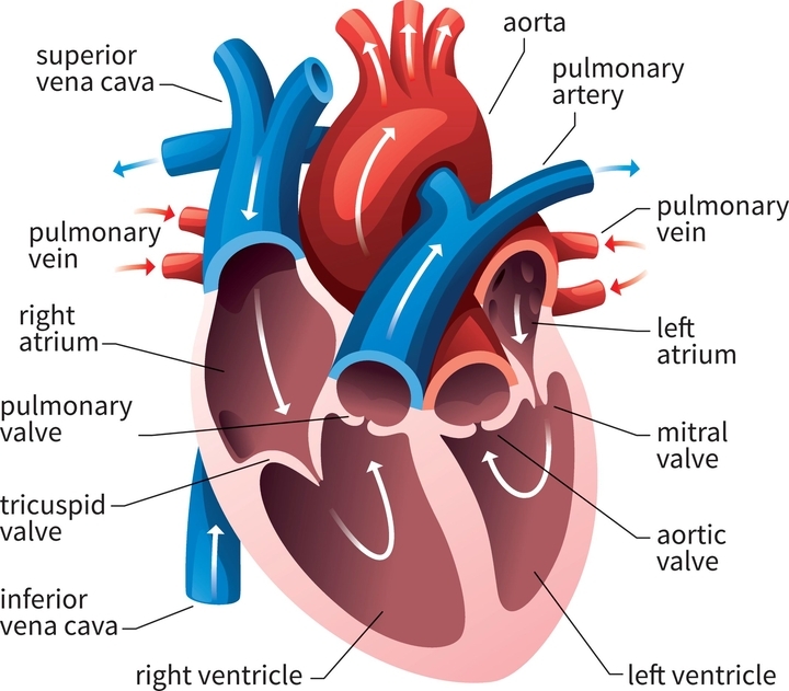
The heart is a muscular organ located directly between the lungs in the chest cavity. It is bordered by the pericardium and is placed in the thoracic area, somewhat to the left. The human heart has four chambers: two upper chambers called atria (plural: atrium) and two lower chambers called ventricles.
Wall: Three layers make up the heart wall. The outside layer of the heart wall is called the epicardium, the intermediate (and muscular) layer is called the myocardium, and the innermost layer is called the endocardium.
Chambers: The heart is divided into four chambers: the right and left atria, as well as the right and left ventricles. They make up the interior cavity of the heart. The four chambers are crucial in circulation. The atria take in blood from the veins, whereas the ventricles expel blood from the heart. The ventricle’s myocardial layers are thicker than those of the atria because they must be stronger to execute this pumping role.
Blood Vessels
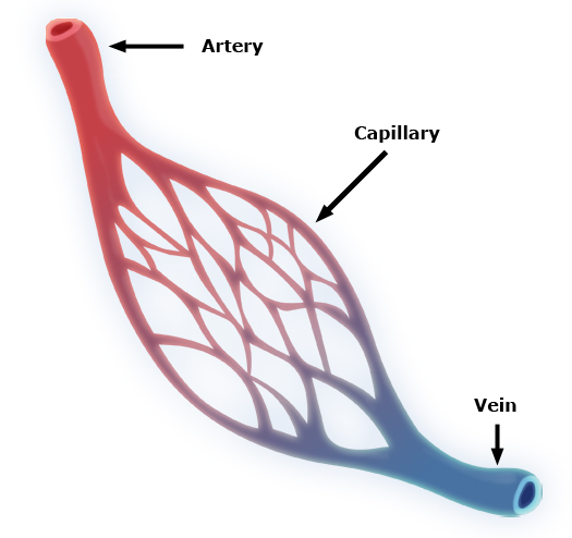
Arteries: Arteries carry blood away from the heart. The artery walls are divided into three layers: tunica intima (inner), tunica media (middle), and tunica externa (outer). The middle layer is usually the thickest. It is made up of smooth muscle that changes the size of the artery to regulate blood flow. Arterioles are divided into three types. They become smaller as they move away from the heart.
Veins: Blood returns to the heart via veins. Blood moves from capillaries to venules (the smallest veins). As the blood draws closer to the heart, the veins enlarge. Veins, like arteries, have three layers of wall: tunica intima, tunica media, and tunica externa. There are some key differences between arteries and veins:
- The walls of veins have less smooth muscle and connective tissue.
- Vein walls are thinner than artery walls.
- Veins have lower pressure and can accommodate more blood than arteries.
At any given time, the veins contain around 70% of the body’s total blood supply.
Blood
Blood transports almost everything in the human body such as hormones, nutrition, oxygen, antibodies, and other vital substances. There are 4 important components of human blood:
- Plasma
- Red Blood Cells (RBC)
- White Blood Cells (WBCs)
- Platelets
Lymphatic System
In the human circulatory system, another body fluid is called lymph. It is also known as tissue fluid. It is produced by the lymphatic system, which is made up of interconnected organs, nodes, and ducts. Lymph is a colorless fluid composed of salts, proteins, and water that transports and circulates digested food and fat between cells in tissues. In contrast to the circulatory system, lymph flows passively via a network of vessels rather than being pumped.
Features of Human Circulatory System
The following are the critical features of the circulatory system:
- The circulatory system of the human body is made up of blood, the heart, blood arteries, and lymph.
- The human circulatory system circulates blood in two loops (double circulation), one for oxygenated blood and the other for deoxygenated blood.
- There are four chambers in the human heart: two ventricles and two auricles.
- The human circulatory system is comprised of a network of blood vessels that spans the entire body. These are made up of arteries, veins, and capillaries.
- Blood vessels’ principal job is to transmit oxygenated blood and nutrients to all regions of the body. It is also in charge of collecting metabolic wastes for expulsion from the organism.
- Most circulatory system diagrams do not show the entire length of the system. In theory, if a human’s veins, arteries, and capillaries were put end to end, they would reach a total distance of 1,00,000 kilometers (approximately eight times the diameter of the Earth).
Functions of Human Circulatory System
The circulatory system’s primary function is to deliver oxygen throughout the body. The following are the other essential activities of the human circulatory system:
- It aids in the maintenance of all organ systems.
- It is responsible for transporting blood, nutrients, oxygen, carbon dioxide, and hormones throughout the body.
- It defends cells from infections.
- It serves as a bridge for cell-to-cell communication.
- Blood-borne chemicals aid in the regeneration of injured tissue.
Human Circulatory System: Working
The lungs’ thin membranes absorb oxygen as it is inhaled, allowing oxygen to enter the bloodstream. The body produces carbon dioxide as it utilizes oxygen and processes nutrients, which your lungs expel as you exhale. The circulatory system functions as a result of continual pressure from the heart and valves located throughout the body. This pressure keeps veins carrying blood to the heart and arteries transporting blood away from the heart.
There are three types of circulation that occur on a regular basis in the body:
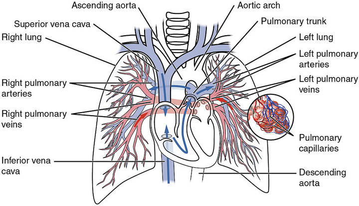
- Pulmonary Circulation: This portion of the cycle transports oxygen-depleted blood from the heart to the lungs and then back to the heart.
- Systematic Circulation: This is the section that transports oxygenated blood from the heart to various regions of the body.
- Coronary Circulation: This form of circulation supplies oxygenated blood to the heart, allowing it to operate effectively.
Course of circulation:
Along with the closed circulatory system, mammals have double circulation which means blood has to cross two times from the heart before circulating throughout the body. The right atrium receives impure blood from the body which goes into the right ventricle. From here the blood went into the pulmonary artery which sends it to the lung for purification. After purification, it is collected by the pulmonary vein which brings it back to the heart in the left atrium. From the atrium, it vents into the left ventricle. Now, this purified blood goes into the aorta for different organs of the body. This circulation is done in a cardiac cycle.
Cardiac cycle:
- The cardiac cycle is controlled by two pacemakers in the heart:
- The sino-atrial node (SA node) is located in the top wall of the right atrium, also known as the heart of the heart.
- The atrioventricular node (AV node) is located between the right atrium and ventricle.
- Both pacemakers are types of nervous tissue.
Blood Pressure:
- The force per unit area is exerted on the walls of the blood-carrying tube by blood called blood pressure.
- The normal rating of blood pressure in a healthy person is 120/80 mm Hg.
- Blood pressure is high in tubes that carry blood to the body parts (systolic pressure) and low in tubes that carry blood to the heart (diastolic pressure).
Wireless Artificial Pacemaker:
- When the SA node fails or is injured, cardiac impulses are not generated.
- For this, we use a wireless pacemaker that regulates the heart by wireless pulses of ultrasound from outside the organ.
- It is beneficial over conventional pacemakers as the leads can fail and require additional surgery to replace them.
Cardiovascular Diseases
The following are the commonly seen Cardiovascular Diseases:
1) Arteriosclerosis:
It is the thickening, hardness, or lack of flexibility of the arteries caused by plaque and calcification formation in their walls. The development of fatty plaques, cholesterol, and other chemicals in and on the arterial walls is the reason behind Arteriosclerosis.
2) Atherosclerosis:
Deposition of cholesterol in the walls of arteries due to which they become narrow and hinder the blood flow to them.
3) Heart Attack:
A heart attack occurs when one or more of your coronary arteries become blocked. Over time, a coronary artery can narrow from the buildup of various substances, including cholesterol (Atherosclerosis).
Human Circulatory System: Important Points
Below are some important facts related to the Human Circulatory System. Aspirants are advised to go through these points in order to be better prepared for the examination.
- The biological term for the heart is ‘Cardio’.
- The heart remains safe in the pericardial membrane.
- Its weight is approximately 300 grams for males and 250 gms for females.
- The human heart is a four-chamber heart.
- In one cycle, the heart pumps 70ml of blood.
- The major function of the heart is the pumping of blood/circulation of blood.
- The heartbeat of a normal human being is 72 beats/minute.
- The heartbeat of the shrew is a maximum of 800 beats per minute.
- It contains the right atrium and a left atrium in the anterior part.
- There is a tricuspid valve between the right atrium and the right ventricle.
- There is a bicuspid valve between the left atrium and left ventricle.
- It contains a right ventricle and a left ventricle persists on the posterior side.
- It is a pumping organ that works in a rhythmic cyclic manner with systole (shrinkage for .3sec) and diastole (expansion for .5 sec).
- A heartbeat lasts for 0.8 seconds and consists of both of these.
- A vein is a vessel that carries blood from the body to the heart.
- The vein contains impure blood i.e. carbon dioxide mixed blood.
- The pulmonary vein is the exception which always carries pure blood.
- The pulmonary vein carries the blood from the lungs to the left atrium.
- The artery is the vessel that carries the blood from the heart toward the body.
- Artery contains pure blood i.e. oxygen mixed blood.
- The exception is the pulmonary artery, which always transports dirty or deoxygenated blood.
- The pulmonary artery carries the blood from the right ventricle to the lungs.
- In the right part of the heart, there remains impure blood i.e. carbon dioxide mixed blood and in the left part of the heart, there remains pure blood i.e. oxygen mixed blood.
- The artery carrying blood to the muscles of the heart is called the coronary artery. Any type of hindrance in it causes a heart attack.
All the Best